Alzheimer’s disease


What is the difference between Dementia and Alzheimer’s disease?

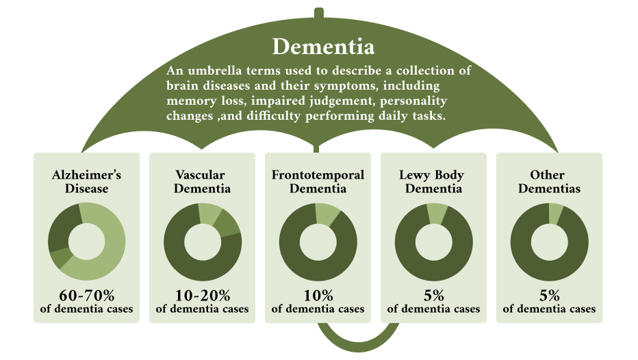
Dementia is not a specific disease or a specific diagnosis. It is a group of symptoms resulting from an affliction of the brain from many diverse causes. It is a general term for any slow and chronic decline in mental ability with short term memory loss, changes in thinking and behavior that affects daily life and has a variety of causes. Dementia shows a slow progressive decline with inability to learn or retain information, inability to reason and a decline in cognitive  function, such as thinking, reading, retaining information, changes in language and speech, trouble planning and loss of organizational skills.These changes happen very slowly over a period of years.

A good analogy to the term dementia is “fever”. Fever refers to an elevated temperature, indicating that a person is sick. But it does give any information about what is causing the sickness. In the same way, dementia means there is something wrong with the brain, but it does not provide any information about what is causing the memory or cognitive issues. Dementia is like a symptom or symptoms, indicating that there is  problem in the functioning of the brain.

What is the most common cause of dementia?

Alzheimer’s disease accounts for 60-80 % of all cases of dementia and is the most common cause of dementia.

What are the ten early signs and symptoms of Alzheimer’s disease?

https://alz.org/10-signs-symptoms-alzheimers-dementia.asp?gclid=EAIaIQobChMIhqSekMCh2QIVQoezCh0tZAQNEAAYASAAEgIcbfD_BwE

Can the diagnosis of Alzheimer’s disease be confirmed by a blood test, brain imaging studies by MRI or other special brain scans or any other test?

Alzheimer’s disease cannot be confirmed by any known tests. The cause remains a mystery and tests are done to rule out other causes of brain dysfunction or the slow altered mental state that has many causes that include, alcohol, narcotics, sedatives, vascular dementia or strokes as well as many other reversible and irreversible diagnoses. The unfortunate journey of the aging patient afflicted by Alzheimer’s disease is further interrupted by multiple medications, multiple health professionals, multiple hospital admissions, multiple residence in various assisted living facilities and specialty assisted living facility and long term facilities and a cascading spiral towards an unfortunate death with suffering along the way and the tragedy is that this can be avoided!

Alzheimer’s disease can be diagnosed with complete accuracy only after death, upon autopsy when microscopic examination of the brain with special stains reveals the characteristic Amyloid plaques and tangles.

When all known causes of dementia are excluded, a clinical diagnosis of Alzheimer’s disease is made.

How is a clinical diagnosis of Alzheimer’s disease made?

A clinical diagnosis of Alzheimer’s disease is made by proceeding with the following:

  1. Detailed history from close family members indicating the chronic changes in mental state that they have noticed, such as short term memory loss with the patient unable to recall things or converstaions that have happen recently and s low changes in behavior and personality and notes taken from multiple sources indicating concerns in mental changes in the patient.
  2. A complete physical examination and a complete blood work that includes vitamin B12 level and Thyroid function tests.
  3. A non-contrast CAT scan of the head and if indicated a MRI of the brain.

If the above show no significant abnormality, the next step is to perform Cognitive tests.

What cognitive Function tests does your office perform?

Mini-Cog test: this is a simple three minute test that is useful in detecting Alzheimer’s disease and research has shown that it has nearly 83% accuracy.

The first part of the test involves asking the patient to repeat three words from the choices given on the test form. The patient than is asked to draw a clock by placing in the numbers and than ask the patient to set the hands of the clock to 10 past 11.

The patient than is asked to recall the three words and if they are unable to recall the words, there is an indication that there is potentially short term memory issue.

How is the clock drawing test scored?

The simplest scoring method consists of giving one point if the task is completed and zero points if the clock was not completed correctly. However , it is important to look for the inclusion of each number, correctly ordered numbers, two clock hands and drawing the correct time.

Examples of clock drawing by patient’s in our practice with Alzheimer’s disease?

What is the reason that patient’s with Alzheimer’s disease fail in drawing the clock correctly?

The clock drawing test is an excellent tool to screen different cognitive domains. The test detects problems in executive functioning i.e , ability to organize, plan and carry out a set of tasks in an efficient manner. It also includes the ability to self-monitor and control our behavior.There is a loss or impairment in accessing the knowledge of the attributes, features, and the meaning of a clock.

What are the other cognitive tests done at at the office?

  1. GPCOG screening test, further assess brain function and in addition gets further information from family and /or caregivers.
  2. MOCA screening test ( Montreal Cognitive assessment -MoCA) is widely used screening assessment for cognitive impairment. This is a more sensitive screening test for Dementia.

How did they come up with the name Alzheimer’s?

The disease was named  to honor Dr. Alois Alzheimer ( 1864-1951), a German Neuropathologist. In 1901 Dr.Alzheimer closely watched a 51 year old patient, Ms.Auguster Deter who  developed a strange behavior, including short term memory loss. She died in 1906 and a brain autopsy was done and Dr. Alzheimer uses special stain techniques to study the brain tissue under the microscope and identified Amyloid plaques and Neurofibrillary tangles. These two abnormal protein deposits inside and the outside the nerve cells, scattered amongst normal nerve cells lead a a slow degeneration, destruction and ultimately death of the nerve cell and remain the hallmark of this devastating disease.

Dr. Alois Alzheimer died at a young age of 51 ( like all great human beings),  from Rheumatic heart disease. He was a leading specialist in histopathology of the brain in Europe. He died on 19th Dec.1915 and was buried next to his wife in Frankfurt, Germany.

WHAT DOES A NEURON LOOK LIKE AND HOW DOES IT WORK?

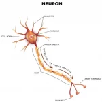
There are nearly 100 Billion neurons or nerve cells in the Brain . A neuron contains three parts, a cell body that directs all cell activity and makes proteins or neurotransmitters, dendrites (that look like branches of a tree), which are short fibers that receive messages from other neurons and relay these messages to the cell body and this passed on to the axon that conducts electrical impulses and transmits information to other neurons, muscles and glands.

How does Alzheimer’s disease affects the brain?

Many molecular and cellular changes take place in the brain of a patient with Alzheimer’s disease. These changes can be observed in brain tissue under the microscope after death. Research continues and the following changes in the brain have been seen:

Amyloid plaques:

Amyloid protein is normally formed in the organs and easily cleared. For reasons that are not known abnormal levels of this naturally occurring protein clump together to form plaques that collect between neurons and interrupt communication and interaction between cells, interfere with transmission of messages, disrupt blood vessels, destroy the local micro neuronal environment, cut of the supplies,engulg and suffocate the cells and strangulate them leading to the ultimate death of the cell and replaced with scar tissue!

Neurofibrillary Tangles

There is another naturally occurring protein, called Tau protein that for unknown reason changes its behavior and accumulates inside the nerve cell. Normal Neurons have microtubules that help transport molecules and nutrients from the cell body to the dendrites and axons. In normal neurons Tau protein binds and strengthens these microtubules. In Alzheimer’s disease there is a change in the structure of Tau protein and they detach from the microtubule and stick to each other leading to formation of thread like structures, called Neurofibrillary tangles and these get entangled together and block the neurons transport system and disrupt the local communicating system.

Dysfunction of Microglia

Microglia are normal cells located throughout the brain and account for 10-15% of all cells found within the brain. They act as the first and main form of active immune defense in the nervous system. They are responsible for protection of the brain from invaders and act as scavengers to get rid of unwanted or invading pathological molecules. For unknown reasons they get chronically activated by a single stimulus e.g., damage to neuron or by  multiple stimuli that lead to chronic activation and inflammation leading to neuronal death.

Research is ongoing about multiple factors playing a role and no definite theory is available to know exactly why there is a slow degeneration of brain cells.

What are the risk factors of Alzheimer’s disease?

Alzheimer’s disease and aging :

Alzheimer’s disease is not a normal part of the aging process, however, with advancing age , the chances of developing the disease increases. At the age of 70 and above ,about 14 % of the population in Unites States have Alzheimer’s disease, after age 65 the disease doubles every 5 years. After age 85, the risk of developing the disease is 50%.

Will I get Alzheimer’s if a parent has the disease?

If your parent or sibling had Alzheimer’s disease, your risk increases by 30%. To clarify this and get a better understanding before panicking it is to understand the risk in the correct context. If you are 65, the risk of being diagnosed with Alzheimer’s is 2% per year, although this also means a 98% chance per year of not developing the disease. A family history of Alzheimer’s raises 2% annual risk  and family history raises the annual risk by 30%, to 2.6 per year. The risk of developing Alzheimer’s with a family history is very small. The risk is more , if the mother had Alzheimer’s compared with the paternal history.

Is there a genetic test that can diagnose Alzheimer’s disease and what is the role of genetics in the disease?

There is no genetic test that can diagnose Alzheimer’s disease. Research is ongoing and the following genes connected to Alzheimer’s disease have been identified.

Researchers have not found a single gene that directly causes the late- form of the disease in which symptoms become apparent after the age of 65 and later. However, one genetic factor- having one form of a apolipoprotein E (APOE) gene on chromosome 19 – does increase a persons risk.

Early-Onset Alzheimer’s disease occurs in person’s 30s to mid-60s and is responsible for about 10% of all cases with Alzheimer’s disease. Early-onset involving families and the other with no significant family history.

Early-onset familial Alzheimer’s disease, or FAD can occur when there is a single gene mutation or change on chromosomes 21,14 and 1. Each of these mutations cause formation of abnormal Amyloid protein deposition in the brain just as in late-onset disease. A child whose biological mother or father carries a genetic mutation of the above mentioned chromosomes has a 50 % chance of inheriting this disease. This of a major breakthrough as this gives an opportunity to observe the Alzheimer’s-related  brain changes that occur in these families long before symptoms of memory loss appear. The deposition of  abnormal Amyloid protein occurs in both early-onset and late-onset of the disease and there is a greater opportunity to test the effectiveness of Amyloid-clearing drugs in early-onset familial Alzheimer’s disease.

Do patient’s with Down syndrome develop beta-amyloid deposition leading to Alzheimer’s disease?

Down syndrome – also known as Trisomy 21- in a condition where a child is born with extra genetic material present in chromosome 21. Autopsy studies have shown that by age 40, the brain of patients with Down syndrome have significant levels of beta-amyloid protein and Tau tangles, the same abnormal deposits that are the hallmark of Alzheimer’s disease.

How are Beta-amyloid plaques and Tau neurofibrillary tangles formed?

https://www.khanacademy.org/science/health-and-medicine/mental-health/dementia-delirium-alzheimers/v/alzheimers-disease-plaques-and-tangles

What is the cause for the formation of Beta amyloid protein and neurofibrillary tangles in Alzheimer’s disease?

In 1906 Dr. Eloisa Alzheimer was the first Physician to identify the amyloid plaques and neurofibrillary tangles and to this day the abnormal amyloid deposition remains the central event in the etiology of Alzheimer’s disease. The unanswered and challenging question that remains a puzzle  is – what is causing this deposition?

There are multiple theories:

  1. Genetic theory- Beta amyloid protein is a naturally occurring protein and the gene for this is located in chromosome 21 and patient’s affected by Down syndrome have a higher incidence of Alzheimer’s disease after age 35. Likewise, a change or mutation of the gene AAP (Amyloid precursor protein) on chromosome 21 leads a to a change in the molecular structure of the amyloid protein and this prevents the local brain structures from removing this protein and accumulation in the brain initiates the slow process of destruction of the neuron.

Can current medications slow down, reverse or prevent the progression of Alzheimer’s disease?

International research has shown no evidence, that any current medications can slow down, reverse or prevent the underlying disease process.

The U.S Food and Drug Administration (FDA) has approved a number of drugs to treat symptoms of Alzheimer’s disease.

The FDA approved the following drugs to treat the symptoms of Alzheimer’s disease.

This group of medications are called- Cholinesterase  inhibitors and their mechanism of action is the same and any one of them can be prescribed.

1.Aricept (Generic name is Donepezil )

2.Exelon ( Generic name is Rezadyne)

3. Razadyne ( Generic name is Galantamine)

The second type of medication is :

1.Namenda ( Generic name is Memantine)

A combination of Aricept and Namenda is available as Namzeric.

How do these FDA approved drugs to treat symptoms of Alzheimer’s disease work?

The brain cells make chemical proteins that act as messengers and transmit information and coordination to allow brain cells to function, they are called neurotransmitters. according to researchers there are 30-100 different molecules types and with 10 of them doing 99% of the work.

One of the many neurotransmitters that coordinate the complex function of the brain , is Acetylcholine.

The current medications for Alzheimer’s disease prevent the breakdown of this chemical, Acetycholine, and it is postulated that there is an increased availability of this neurotransmitter  in the remaining cells of the brain that are not dead, dying or in the process of a slow death from deposition of the abnormal protein and this increased neurotransmitter in the brain is  approved to treat some of the symptoms of the disease.

What evidence is there that these medications work?

The current medications for Alzheimer’s do not alter, delay, halt or reverse the deposition of the abnormal protein, it is believed that they help with some of the many symptoms of the disease, while the disease continues to progress. The drug insert from the company that makes these drugs reads as follows- ” there is no evidence that these medications delay or prevent the progression of the underlying disease process”.

These medications may treat some of the many symptoms of the disease and research results usually find that the effect in most people is small to non- existent.

In our practice we discuss the pros and cons of therapy, including the possible lack of benefit, the potential for side effects, the cost and interaction with other drugs and allow the patient and their families to make their own value judgments as to whether to proceed  with pharmacotherapy.

What are the common side effects of these medications?

Nausea, vomiting, diarrhea , loss of appetite, weight loss, insomnia, muscle cramps

Patient’s with Alzheimer’s  have increasing difficulty communicating with others about their experience or have difficulty in interpreting their symptoms. As a result they may be unable to report symptoms.

                    Acute Delirium in the Elderly


A brief summary.

Acute delirium in the elderly is a medical emergency and is due to acute brain failure.

Acute delirium is not a specific disease but the reaction of an aging brain due to an acute medical illness, hospitalization, inappropriate medication, or a metabolic problem e.g. low sodium level in the blood or a combination of multiple factors leading the brain to show signs of brain failure. There  is usually not a single factor causing the problem. In most cases the acute change in mental state is from problems outside the brain.

It it is a frequent and serious complication of hospitalization. Its prevalence in the community is 1-2 % and after hospitalization the incidence ranges from 14-56 % and in the Intensive care unit (ICU) the incidence can reach 70-87%.

Neuroimaging of the brain with CAT scan and MRI of brain of normal elderly patients has shown atrophy of the brain, ventricular dilatation and many other changes related primarily to the normal process of aging. These imaging changes reflect a state of increased vulnerability of the brain to any insult and the brain is unable to cope with an infection or inappropriate medications, medical illness, surgery, anesthesia etc.

The disease starts acutely and occurs over hours and days. The clinical presentation in a hospitalized patient varies and has a wide variety of presentation and can present as acute confusion, lethargy, agitation, visual hallucinations and a combination of multiple symptoms that fluctuate over the course of the day and are worse at night and at the time of interaction with the patient, there may be no symptoms.

There is a no single strategy or an established plan of care in the management of the disease in a hospitalized patient. There is an acute need to develop an evaluation and management protocol and process to address one of the most costly geriatric emergencies in our community.

Acute delirium does not occur in all elderly patients, there are multiple risk factors that make the patient vulnerable to delirium. These risk factors may be modifiable or be completely or partially modifiable or be non-modifiable and this will be discussed below. The key to understanding the pathophysiological processes is to aggressively address these issues in order to make a meaningful difference in the life of our patients.

There is no single strategy or process


What are the symptoms or clinical features of delirium?

Acute Delirium is of sudden onset and usually occurs over hours or days. It is a medical emergency. Signs and symptoms fluctuate throughout the day, and there may be periods where the patient has no symptoms.

The main symptoms of delirium include:

– Clouding of consciousness with reduced awareness of the environment.

– Difficulty maintaining or shifting attention

-Disorientation to time, place or person

– Visual hallucinations

What are the techniques for diagnosis?

The following features must be present for a diagnosis of delirium:

1.Disturbance in attention ( reduced ability to remain focussed), and awareness ( reduced orientation to the environment).

– Attention is assessed by asking patient to name the months of the year backwards.

– Awareness is checked by asking the patient their age, date of birth, place( name of the hospital) and current year.

2. The disturbance develops over a short period of time (usually hours or days), represents an acute change from baseline and fluctuates in severity with possible periods of lucidity during the course of a day.

– Establishing this requires close communication amongst staff or family members and information about previous cognitive status.

3. Disturbances in cognition e.g. fluctuating confusion, memory and language deficits, disorientation and confirming that there is no previous existing dementia, psychosis or another neurodegenerative disorder.

4. The most important point is to make sure that there is evidence from the patient’s history, physical exam, laboratory findings and appropriate investigations, that the delirium is a direct physiological consequence of another medical condition, substance intoxication or withdrawal, administration of an inappropriate medications. The causes are usually multifactorial.

What are the types of delirium?

1. Hypoactive form – Predominant symptoms are drowsiness and inattention. A study which examined 805 consecutive acute medical admissions reported that 75% of cases of acute delirium were missed by the admitting team . There are many reasons why hypolert delirium is missed :

– Persons too withdrawn to alter a care provider, particularly if isolated without family or care giver.

– The condition fluctuates and periods of near- normality may coincide with a clinician’s assessment.

– Establishing the diagnosis requires longitudinal overview, to capture the shift from baseline, combines with careful assesment.

2. Hyperalert form- patients are agitated and account for 20 % of al cases of delirium.

3. Mixed form- a combination of the above .


Investigations to consider when assessing a patient with suspected delirium.

In majority of cases the primary problem in delirium is outside the central nervous system and the aging brain is reacting to the stress.

The choice of test will depend on the individual patient. Since there are multiple factors causing delirium, the following process is likely to be helpful in reaching  diagnosis.

The first step is Medication review in order to identify those that are contributory or causative.

The choice of tests depend on the individual patient.

_ Complete blood count (CBC)- an elevated white blood cell found could indicate an infection and anemia may be a clue to an underlying acute medical illness.

Basic metabolic profile (BMP)- the following changes can contribute to delirium

– a low or high sodium level- due to any cause

– a high calcium level from any cause needs to be addressed.

– raised BUN/creatinine or GFR- indicating kidney damage may contribute to the problem

– A low or a very high blood sugar level

_. Liver function tests- advanced liver disease causes delirium, particulalry raised liver enzymes

_Thyroid function tests- for hypothyroidism or hyperthyroidism need to be ruled out.

_ Urine analysis  and urine culture – to detect urinary tract infection and is one of the important cause or a  contributory factor for the development of acute delirium urinary tract infection

_ Blood cultures – for bacteria in the blood stream

_Cardiac enzymes and EKG- silent myocardial infarction can contribute to precipitating acute delirium in a hospitalized patient.

_chest x ray – pulmonary infections are a common cause for precipitating an episode of acute delirium.

_ Arterial blood gases – for low oxygen or high carbon dioxide levels.

_ Computed tomography of head (CAT scan)-which may show a primary neurological problem as the cause of acute delirium.


coming soon

The Center For Elder Care provides specialized care for the unique needs of older adults. Our clinicians are experts in managing geriatric patients and specialize in management of multiple, complex, acute, and chronic medical conditions. 

A few of the unique clinical challenges in round-the-clock care of the elderly include: 

Alzheimers Disease and Dementia

These patients are vulnerable and require close monitoring. Our staff provides sympathetic support to our patients and their families.

www.alz.org

Falls, Gait, and Balance Problems in Older Adults

Each year 1 in 3 community dwelling individuals above the age of 65 fall each year in the United States. These problems are due to multiple factors and our team specializes in a global approach to fall prevention based on gait and balance assessment, medication review, etc. These interventions can prevent falls by up to 60 – 70%. We established one of the first evidence-based fall prevention program based on scientific evidence, international research, and are following the CDC (Center for Disease Control) model for fall prevention. We established a program called “Balance for Life”, offering exercises for an hour three days a week in fourteen churches in North Alabama. This program is offered to all members of the community and is free of cost. Visit www.centerforaging.net for more information. www.cdc.gov/steadi

Polypharmacy

Polypharmacy is the use of four or more medications, prescribed or over-the-counter medications, by an older adult. The daily use of these medications is necessary and appropriate, however, it is impossible to know or predict the side effects of a combination of drugs due to drug interaction and drug to food interaction. It becomes nearly impossible to differentiate between the side effect of a medication from the symptom of a disease process. Our clinical staff continues to learn the art of skillful management of medications from our patients.

The Beer’s list is a list of potentially inappropriate medication for older adults over the age of 65.

www.americangeriatrics.org/files/documents/beers/PrintableBeersPocketCard.pdf

Depression

Patients face many challenges as they navigate through the aging process. The aging process is accompanied by loss of social support systems due to loss of spouse or siblings, retirement, relocation of residence to a new community, disruption in social and family relationships. Our staff screen all our patients with an annual PHQ-9 Depression Scale and/or the Geriatric Depression Scale. We offer support to the family members to alert them about subtle symptoms of depression that may creep in older adults.

Acute delirium in the elderly

Acute delirium is the most common complication of hospitalization in the elderly population. The incidence of acute delirium during a hospital stay varies from 14% to 56% and 70% to 87% in the ICE. Development of delirium depends on a complex interaction of multiple risk factors. More than 50% to 60% of the disease entity can be prevented by appropriate intervention. The mortality of the disease entity rates from 10% to 65 %. We use the CAM scale ( Confusion Assessment Scale) to diagnose the disease and treat based on evidence based practice of medicine.